Inducing sterile pyramidal neuronal death in mice to model distinct aspects of gray matter encephalitis
Abstract
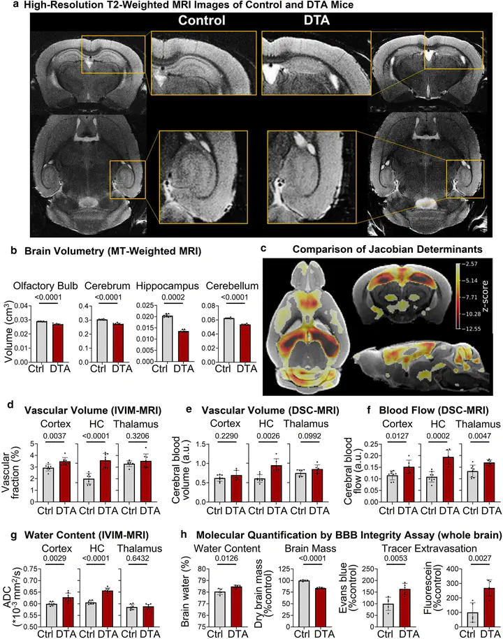
Up to one person in a population of 10,000 is diagnosed once in lifetime with an encephalitis, in 50–70% of unknown origin. Recognized causes amount to 20–50% viral infections. Approximately one third of affected subjects develops moderate and severe subsequent damage. Several neurotropic viruses can directly infect pyramidal neurons and induce neuronal death in cortex and hippocampus. The resulting encephalitic syndromes are frequently associated with cognitive deterioration and dementia, but involve numerous parallel and downstream cellular and molecular events that make the interpretation of direct consequences of sudden pyramidal neuronal loss difficult. This, however, would be pivotal for understanding how neuroinflammatory processes initiate the development of neurodegeneration, and thus for targeted prophylactic and therapeutic interventions. Here we utilized adult male NexCreERT2xRosa26-eGFP-DTA (= ‘DTA’) mice for the induction of a sterile encephalitis by diphtheria toxin-mediated ablation of cortical and hippocampal pyramidal neurons which also recruits immune cells into gray matter. We report multifaceted aftereffects of this defined process, including the expected pathology of classical hippocampal behaviors, evaluated in Morris water maze, but also of (pre)frontal circuit function, assessed by prepulse inhibition. Importantly, we modelled in encephalitis mice novel translationally relevant sequelae, namely altered social interaction/cognition, accompanied by compromised thermoreaction to social stimuli as convenient readout of parallel autonomic nervous system (dys)function. High resolution magnetic resonance imaging disclosed distinct abnormalities in brain dimensions, including cortical and hippocampal layering, as well as of cerebral blood flow and volume. Fluorescent tracer injection, immunohistochemistry and brain flow cytometry revealed persistent blood–brain-barrier perturbance and chronic brain inflammation. Surprisingly, blood flow cytometry showed no abnormalities in circulating major immune cell subsets and plasma high-mobility group box 1 (HMGB1) as proinflammatory marker remained unchanged. The present experimental work, analyzing multidimensional outcomes of direct pyramidal neuronal loss, will open new avenues for urgently needed encephalitis research.
Rakshit Dadarwal
Neuroimaging Researcher
My research focuses on the acquisition and processing of multi-contrast magnetic resonance imaging (MRI) data. I am particularly interested in imaging myelin and iron in the central nervous system using diffusion weighted imaging (DWI), quantitative susceptibility mapping (QSM), magnetisation transfer imaging and T1 and T2 relaxometry.